Featured Publications
Two-photon polymerization for production of human iPSC-derived retinal cell grafts
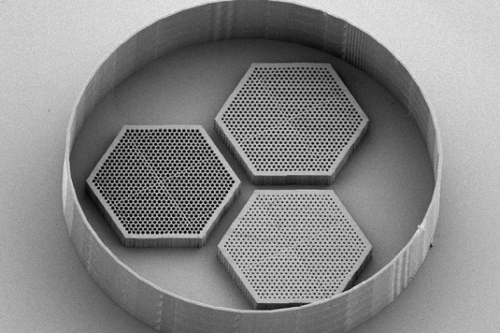
Cell replacement therapy is an important goal for investigators aiming to restore neural function to those suffering from neurodegenerative disease. Cell delivery scaffolds are frequently necessary for the success of such treatments, but traditional biomaterials often fail to facilitate the neuronal orientation and close packing needed to recapitulate the in vivo environment. Here, we use two-photon polymerization to create prototype cell scaffolds with densely packed vertical pores for photoreceptor cell loading and small, interconnected horizontal pores for nutrient diffusion. This study offers a thorough characterization of how two-photon polymerization parameters affect final structural outcomes and printing time. Our findings demonstrate the feasibility of using two-photon polymerization to create scaffolds that can align neuronal cells in 3D and are large enough to be used for transplantation. In future work, these scaffolds could comprise biodegradable materials with tunable microstructure, elastic modulus and degradation time; a significant step towards a promising treatment option for those suffering from late-stage neurodegeneration, including retinal degenerative blindness.
Mechanical properties of murine and porcine ocular tissues in compression
Sub-retinal implantation of foreign materials is becoming an increasingly common feature of novel therapies for retinal dysfunction. The ultimate compatibility of implants depends not only on their in vitro chemical compatibility, but also on how well the mechanical properties of the material match those of the native tissue. In order to optimize the mechanical properties of retinal implants, the mechanical properties of the mammalian retina itself must be carefully characterized. In this study, the compressive moduli of eye tissues, especially the retina, were probed using a dynamic mechanical analysis instrument in static mode. The retinal compressive modulus was lower than that of the sclera or cornea, but higher than that of the RPE and choroid. Compressive modulus remained relatively stable with age. Conversely, apparent retinal softening occurred at an early age in mice with inherited retinal degeneration. Compressive modulus is an important consideration for the design of retinal implants. Polymer scaffolds with moduli that are substantially different than that of the native tissue in which they will ultimately reside will be less likely to aid in the differentiation and development of the appropriate cell types in vitro and will have reduced biocompatibility in vivo.
Neuronal differentiation of induced pluripotent stem cells on surfactant templated chitosan hydrogels
The development of effective tissue engineering materials requires careful consideration of several properties beyond biocompatibility, including permeability and mechanical stiffness. While surfactant templating has been used for over a decade to control the physical properties of photopolymer materials, the potential benefit of this technique with regard to biomaterials has yet to be fully explored. Herein we demonstrate that surfactant templating can be used to tune the water uptake and compressive modulus of photo-cross-linked chitosan hydrogels. Interestingly, templating with quaternary ammonium surfactants also hedges against property fluctuations that occur with changing pH. Further, we demonstrate that, after adequate surfactant removal, these materials are nontoxic, support the attachment of induced pluripotent stem cells and facilitate stem cell differentiation to neuronal phenotypes. These results demonstrate the utility of surfactant templating for optimizing the properties of biomaterials intended for a variety of applications, including retinal regeneration.
Development of high-resolution three-dimensional-printed extracellular matrix scaffolds and their compatibility with pluripotent stem cells and early retinal cells
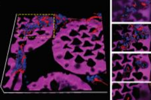
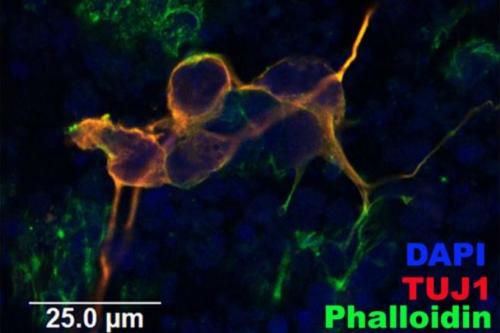
Widely used approaches for retinal disease modeling and in vitro therapeutic testing can be augmented by using tissue-engineered scaffolds with a precise 3-dimensional structure. However, the materials currently used for these scaffolds are poorly matched to the biochemical and mechanical properties of the in vivo retina. Here, we create biopolymer-based scaffolds with a structure that is amenable to retinal tissue engineering and modeling. Methods: Optimal two-photon polymerization (TPP) settings, including laser power and scanning speed, are identified for 4 methacrylated biopolymer formulations: collagen, gelatin, hyaluronic acid (HA), and a 50/50 mixture of gelatin/HA, each with methylene blue as a photoinitiator. For select formulations, fabrication accuracy and swelling are determined and biocompatibility is evaluated by using human induced pluripotent stem cells and rat postnatal retinal cells. Results: TPP is feasible for each biopolymer formulation, but it is the most reliable for mixtures containing gelatin and the least reliable for HA alone. The mean size of microscaffold pores is within several microns of the intended value but the overall structure size is several times greater than the modeled volume. The addition of HA to gelatin scaffolds increases cell viability and promotes neuronal phenotype, including Tuj-1 expression and characteristic morphology.